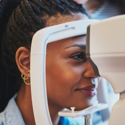
Digital Retinal Photography
The clarity of a high-resolution image of your eye is a game-changer.
Digital Retinal Photography Makes it Easier to Diagnose Eye Disease
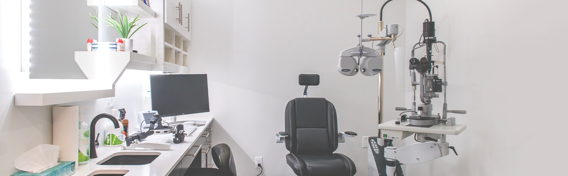
We’ve heavily invested in advanced technology to provide you and your family with next tier eye care. Digital retinal photography allows us to view the back of your eye in extraordinary detail.
Because of this, we can detect signs of many eye diseases in their infancy, enabling us to address these concerns early. When it comes to eye diseases, early detection and treatment is key to preventing vision loss.
Digital retinal images are non-invasive and instant, eliminating wait time. We’re able to immediately use the information gained from these photographs to infer problems, make assessments, and provide us insights into the state of your eye health.
As you can imagine, the ability to see—in ultra high-resolution—what is happening in the eye facilitates easier and more accurate diagnoses of many eye diseases. This tool is Immensely effective for the detection of many eye diseases, including:
Our optometrist will go over the results during your next eye exam and indicate what they are seeing and where.
Often, we see the signs of high blood pressure or diabetes before they are even on our patients’ radar.
What Is Retinal Fundus Photography, and How Does It Work?
Digital retinal photography is also known as digital fundus photography. Fundus photography simply refers to photographing the back of the eye.
Your retina consists of 10 layers. Fundus photography provides a top-down view through these layers to the back of your eye. Using fundus photography, we can detect and monitor the development of many eye diseases.
Zeiss Clarus 500 Widefield Fundus Camera
We are excited to introduce the new Zeiss Clarus 500 Widefield Fundus Camera to NTOC! This innovative tool takes 210° panoramic photographs of the fundus, allowing our doctors to see down to 7 microns and capture retinal changes in the earliest stages.
There are only a few of these cameras in Canada. Your eye health means so much to us that we had to invest in one.